
 说明书
说明书 MSDS
MSDS产品描述:Rabbit polyclonal antibody to HIF1 alpha免疫原:KLH-conjugated synthetic peptide encompassing a sequence within the center region of human HIF1 alpha. The exact sequence is proprietary.纯化方式:The antibody was purified by immunogen affinity chromatography.克隆类型:Polyclonal产品形式:Liquid in 0.42% Potassium phosphate, 0.87% Sodium chloride, pH 7.3, 30% glycerol, and 0.01% sodium azide.稀释比:WB (1/500 - 1/1000), IH (1/50 - 1/100), IF/IC (1/50 - 1/200)基因名称:HIF1A相关名称:BHLHE78; MOP1; PASD8; Hypoxia-inducible factor 1-alpha; HIF-1-alpha; HIF1-alpha; ARNT-interacting protein; Basic-helix-loop-helix-PAS protein MOP1; Class E basic helix-loop-helix protein 78; bHLHe78; Member of PAS protein 1; PAS domain-containing protein 8
基因编号(人):
3091;
基因编号(小鼠):
15251;
基因编号(大鼠):
29560;
蛋白编号(人):
Q16665;
蛋白编号(小鼠):
Q61221;
蛋白编号(大鼠):
O35800;
储存效期:Shipped at 4°C. Upon delivery aliquot and store at -20°C for one year. Avoid freeze/thaw cycles.
-
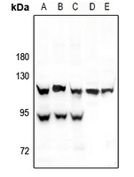 Western blot analysis of HIF1 alpha expression in HEK293T (A), A549 (B), MCF7 (C), H9C2 (D), SP20 (E) whole cell lysates. (Predicted band size: 92 kD; Observed band size: 120; 93 kD)
 -
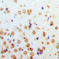 Immunohistochemical analysis of HIF1 alpha staining in human brain formalin fixed paraffin embedded tissue section. The section was pre-treated using heat mediated antigen retrieval with sodium citrate buffer (pH 6.0). The section was then incubated with the antibody at room temperature and detected using an HRP conjugated compact polymer system. DAB was used as the chromogen. The section was then counterstained with haematoxylin and mounted with DPX.
 -
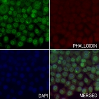 Immunofluorescent analysis of HIF1 alpha staining in LO2 cells. Formalin-fixed cells were permeabilized with 0.1% Triton X-100 in TBS for 5-10 minutes and blocked with 3% BSA-PBS for 30 minutes at room temperature. Cells were probed with the primary antibody in 3% BSA-PBS and incubated overnight at 4 °C in a hidified chamber. Cells were washed with PBST and incubated with a AF488-conjugated secondary antibody (green) in PBS at room temperature in the dark. Phalloidin - AF594 was used to stain Actin filaments (red). DAPI was used to stain the cell nuclei (blue).
Effect of αvβ3 Blockade Against Acute Lung Injury Induced by Influenza A Virus and Its Mechanism
Zi-Su-Zi decoction improves airway hyperresponsiveness in cough-variant asthma rat model through PI3K/AKT1/mTOR, JAK2/STAT3 and HIF-1α/NF-κB signaling pathways
Regulation of Lactate Accumulation in Bovine Mammary Epithelial Cells by Lps-Induced Hif-1α/Mct1 Pathway