
 说明书
说明书 MSDS
MSDS产品描述:Rabbit polyclonal antibody to Involucrin免疫原:KLH-conjugated synthetic peptide encompassing a sequence within the C-term region of human Involucrin. The exact sequence is proprietary.纯化方式:The antibody was purified by immunogen affinity chromatography.克隆类型:Polyclonal产品形式:Liquid in 0.42% Potassium phosphate, 0.87% Sodium chloride, pH 7.3, 30% glycerol, and 0.01% sodium azide.稀释比:WB (1/500 - 1/1000), IH (1/100 - 1/200), IF/IC (1/100 - 1/500)基因名称:IVL相关名称:Involucrin
基因编号(人):
3713;
蛋白编号(人):
P07476;
储存效期:Shipped at 4°C. Upon delivery aliquot and store at -20°C for one year. Avoid freeze/thaw cycles.
-
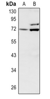 Western blot analysis of Involucrin expression in HEK293T (A), H1792 (B) whole cell lysates. (Predicted band size: 68 kD; Observed band size: 68 kD)
 -
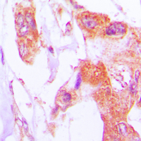 Immunohistochemical analysis of Involucrin staining in human lung cancer formalin fixed paraffin embedded tissue section. The section was pre-treated using heat mediated antigen retrieval with sodium citrate buffer (pH 6.0). The section was then incubated with the antibody at room temperature and detected using an HRP conjugated compact polymer system. DAB was used as the chromogen. The section was then counterstained with haematoxylin and mounted with DPX.
 -
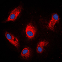 Immunofluorescent analysis of Involucrin staining in MCF7 cells. Formalin-fixed cells were permeabilized with 0.1% Triton X-100 in TBS for 5-10 minutes and blocked with 3% BSA-PBS for 30 minutes at room temperature. Cells were probed with the primary antibody in 3% BSA-PBS and incubated overnight at 4 °C in a humidified chamber. Cells were washed with PBST and incubated with a DyLight 594-conjugated secondary antibody (red) in PBS at room temperature in the dark. DAPI was used to stain the cell nuclei (blue).
Laminin-511 and integrin beta-1 in hair follicle development and basal cell carcinoma formation
Endogenous two-photon fluorescence imaging elucidates metabolic changes related to enhanced glycolysis and glutamine consumption in precancerous epithelial tissues
Notch inhibition allows oncogene-independent generation of iPS cells