
 说明书
说明书 MSDS
MSDS产品描述:Rabbit polyclonal antibody to ERAS免疫原:KLH-conjugated synthetic peptide encompassing a sequence within the C-term region of human ERAS. The exact sequence is proprietary.纯化方式:The antibody was purified by immunogen affinity chromatography.克隆类型:Polyclonal产品形式:Liquid in 0.42% Potassium phosphate, 0.87% Sodium chloride, pH 7.3, 30% glycerol, and 0.01% sodium azide.稀释比:WB (1/500 - 1/1000), IF/IC (1/100 - 1/500)基因名称:ERAS相关名称:HRAS2; HRASP; GTPase ERas; E-Ras; Embryonic stem cell-expressed Ras
基因编号(人):
3266;
基因编号(小鼠):
353283;
蛋白编号(人):
Q7Z444;
蛋白编号(小鼠):
Q7TN89;
储存效期:Shipped at 4°C. Upon delivery aliquot and store at -20°C for one year. Avoid freeze/thaw cycles.
-
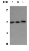 Western blot analysis of ERAS expression in mouse liver (A), mouse testis (B), rat liver (C) whole cell lysates. (Predicted band size: 25 kD; Observed band size: 25 kD)
 -
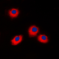 Immunofluorescent analysis of ERAS staining in Jurkat cells. Formalin-fixed cells were permeabilized with 0.1% Triton X-100 in TBS for 5-10 minutes and blocked with 3% BSA-PBS for 30 minutes at room temperature. Cells were probed with the primary antibody in 3% BSA-PBS and incubated overnight at 4 °C in a humidified chamber. Cells were washed with PBST and incubated with a DyLight 594-conjugated secondary antibody (red) in PBS at room temperature in the dark. DAPI was used to stain the cell nuclei (blue).