
 说明书
说明书 MSDS
MSDS产品描述:Recombinant rabbit monoclonal antibody to LDHA免疫原:KLH-conjugated synthetic peptide encompassing a sequence within human LDHA. The exact sequence is proprietary.纯化方式:The antibody was purified by immunogen affinity chromatography.克隆类型:Monoclonal产品形式:Liquid in PBS, pH 7.4, containing 50% glycerol, 0.2% BSA and 0.01% sodium azide.稀释比:WB (1/500 - 1/1000), IH (1/50 - 1/200), IF/IC (1/50 - 1/200)基因名称:LDHA相关名称:L-lactate dehydrogenase A chain; LDH-A; Cell proliferation-inducing gene 19 protein; LDH muscle subunit; LDH-M; Renal carcinoma antigen NY-REN-59
基因编号(人):
3939;
基因编号(小鼠):
16828;
基因编号(大鼠):
24533;
蛋白编号(人):
P00338;
蛋白编号(小鼠):
P06151;
蛋白编号(大鼠):
P04642;
储存效期:Shipped at 4°C. Upon delivery aliquot and store at -20°C for one year. Avoid freeze/thaw cycles.
-
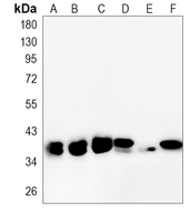 Western blot analysis of LDHA expression in HEK293T (A), Hela (B), Jurkat (C), mouse kidney (D), rat liver (E), rat kidney (F) whole cell lysates. (Predicted band size: 36 kD; Observed band size: 37 kD)
 -
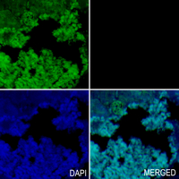 Immunohistochemical analysis of LDHA staining in rat kidney formalin fixed paraffin embedded tissue section. The section was pre-treated using heat mediated antigen retrieval with sodium citrate buffer (pH 6.0). The section was then incubated with the antibody at room temperature and detected using an HRP conjugated compact polymer system. Tyramide-AF488 (green) was used as the chromogen. The section was then counterstained with DAPI (blue).
 -
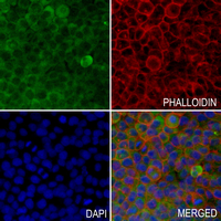 Immunofluorescent analysis of LDHA staining in MCF7 cells. Formalin-fixed cells were permeabilized with 0.1% Triton X-100 in TBS for 5-10 minutes and blocked with 3% BSA-PBS for 30 minutes at room temperature. Cells were probed with the primary antibody in 3% BSA-PBS and incubated overnight at 4 °C in a humidified chamber. Cells were washed with PBST and incubated with an AF488-conjugated secondary antibody (green) in PBS at room temperature in the dark. Phalloidin - AF594 was used to stain Actin filaments (red). DAPI was used to stain the cell nuclei (blue).